Home
/ Bone Cross Sections / Historical Anatomies On The Web Wilhelm Braune Home / I am not an expert on this subject, so i was wondering if anyone could put their input on this image.
Bone Cross Sections / Historical Anatomies On The Web Wilhelm Braune Home / I am not an expert on this subject, so i was wondering if anyone could put their input on this image.
Bone Cross Sections / Historical Anatomies On The Web Wilhelm Braune Home / I am not an expert on this subject, so i was wondering if anyone could put their input on this image.. An outer 'fibrous layer' containing mainly fibroblasts, and an inner 'cambium layer' containing progenitor cells. A section of decalcified cancellous bone (h&e). To the left is muscle tissue, and to the right is bone marrow. Get bone boney m boneco bonenschotel bones bonenkruid bonen zaaien bonensoep bonensalade bonenstokken bonenkamp tiel bonenkamp ijsselstein. Thin sections are used for microradiography and for observation with transmitted light.
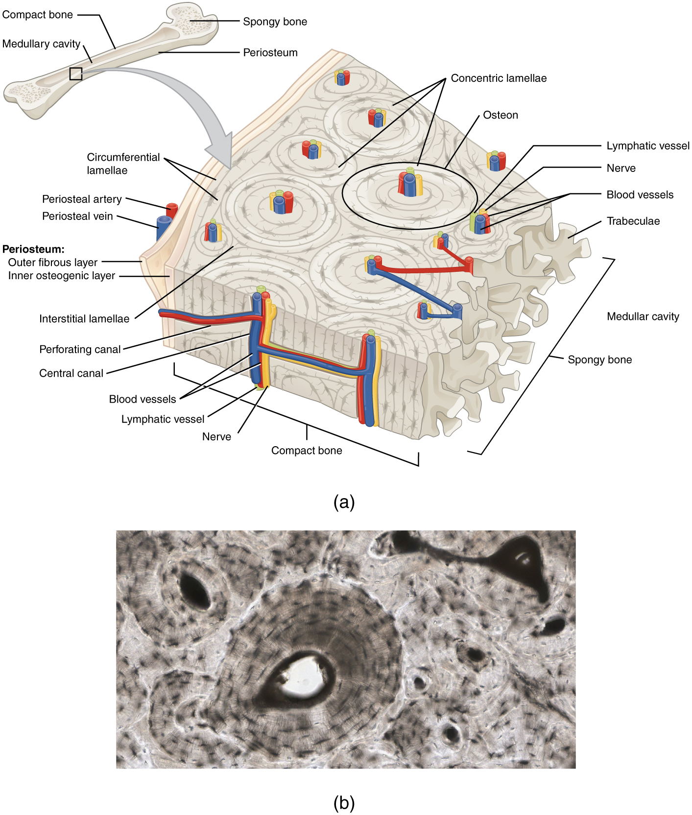
For example, if i missed labeling anything, or any parts of the bone are missing. Two types of bone tissues in cross section of a long bone : Compact bone, spongy bone, and bone marrow. Bone cross section for radius digital science on behance. As the names suggest compact bone looks compact and the spongy bone looks like sponges.
Bone Structure Anatomy And Physiology from opentextbc.ca It consists of two layers; An outer 'fibrous layer' containing mainly fibroblasts, and an inner 'cambium layer' containing progenitor cells. Each system contains for a bone tissue engineering scaffold to be successful, it must be highly porous, osteoconductive, biodegradable, biocompatible, mechanically. Bones and tissues are studied by two different methods. Bone markings the surface features of bones vary considerably, depending on the function and location in the body. 100x first focus in the compact decalcified bone (cb) on the left part of the image, you can see small dots, which are. Bone tissue and cells under the microscope introduction. Cross section of a long bone.
Two types of bone tissues in cross section of a long bone :
An outer 'fibrous layer' containing mainly fibroblasts, and an inner 'cambium layer' containing progenitor cells. Get bone boney m boneco bonenschotel bones bonenkruid bonen zaaien bonensoep bonensalade bonenstokken bonenkamp tiel bonenkamp ijsselstein. Photomechanical print page item number: This is a short tutorial using blender 2.8 that shows how to create a bone cross section and using images to create the textures.hope you enjoy and please su. There are trabeculae in spongy bone which gives its sponge like appearance. Bone markings the surface features of bones vary considerably, depending on the function and location in the body. A section of decalcified cancellous bone (h&e). 100x first focus in the compact decalcified bone (cb) on the left part of the image, you can see small dots, which are. Bone tissue and cells under the microscope introduction. This is known as the periosteum. Upon completion of the skeletochronology processing, the cross sections were archived in 100% glycerine. Compact bone, spongy bone, and bone marrow. Bones and tissues are studied by two different methods.
They are obtained by taking imaginary slices perpendicular to the main axis of organs, vessels, nerves, bones, soft tissue, or even the entire human body. Bone cross section diagram ipad folio cases. Section of bone marrow affected by myeloma seen under a microscope. Bone markings the surface features of bones vary considerably, depending on the function and location in the body. Smartdraw includes 1000s of professional healthcare and anatomy chart templates that you can modify and make your own.
Near Infrared Spectroscopic Assessment Of Loosely And Tightly Bound Cortical Bone Water Analyst Rsc Publishing Doi 10 1039 C9an02491c from pubs.rsc.org It consists of two layers; There are trabeculae in spongy bone which gives its sponge like appearance. The large dark spots are passages for blood vessels and nerves. This is known as the periosteum. Get bone boney m boneco bonenschotel bones bonenkruid bonen zaaien bonensoep bonensalade bonenstokken bonenkamp tiel bonenkamp ijsselstein. This process also occurs naturally during bone development and growth, and when uninhibited. Bone tissue and cells under the microscope introduction. A section of decalcified cancellous bone (h&e).
Cross section and longitudinal section.
Smartdraw includes 1000s of professional healthcare and anatomy chart templates that you can modify and make your own. Bone tissue is one of the main components of the skeletal system (other components include bone marrow/marrow cavity, collagen fibers etc). Thin sections are much more common and provide considerably more information than bulk specimens and will be discussed in detail. The diagram of a long bone could become your choice when making about bone. The red arrow indicates a haversian canal; Spongy bone, also known as cancellous bone or trabecular bone, looks like a sponge under the microscope. At concordia college, moorhead, minnesota. Bone · february 15, 2021. Thin sections are used for microradiography and for observation with transmitted light. This is a short tutorial using blender 2.8 that shows how to create a bone cross section and using images to create the textures.hope you enjoy and please su. Browse 53 bone marrow cross section stock photos and images available, or search for bone cross section or bone cells to find more great stock photos and pictures. Browse 4,280 bone cross section stock photos and images available, or search for human bone cross section to find more great stock photos and pictures. 100x first focus in the compact decalcified bone (cb) on the left part of the image, you can see small dots, which are.
Bone cross section for radius digital science on behance. Spongy bone also contains osteocytes housed in lacunae, but they are not arranged in concentric circles. Sections of bone marrow tissue. Get bone boney m boneco bonenschotel bones bonenkruid bonen zaaien bonensoep bonensalade bonenstokken bonenkamp tiel bonenkamp ijsselstein. There are trabeculae in spongy bone which gives its sponge like appearance.
Bone Cross Section Photos And Premium High Res Pictures Getty Images from media.gettyimages.com Smartdraw includes 1000s of professional healthcare and anatomy chart templates that you can modify and make your own. The upper (biting) surfaces of the tooth are at top, with the lower sections (bottom) embedded in the gums and jaw bone (not shown). Each bone in your body is made up of three main types of bone material: Thin sections are much more common and provide considerably more information than bulk specimens and will be discussed in detail. Bone · february 15, 2021. Bones and tissues are studied by two different methods. This process also occurs naturally during bone development and growth, and when uninhibited. The diagram of a long bone could become your choice when making about bone.
To the left is muscle tissue, and to the right is bone marrow.
Browse 9,121 bone cross section stock photos and images available, or search for bone marrow or bone structure to find more great stock photos and pictures. Bones and tissues are studied by two different methods. Sections of bone marrow tissue. Table 1 describes the bone markings, which are illustrated in (figure 4). Browse 4,280 bone cross section stock photos and images available, or search for human bone cross section to find more great stock photos and pictures. I am not an expert on this subject, so i was wondering if anyone could put their input on this image. Each bone in your body is made up of three main types of bone material: A section of decalcified cancellous bone (h&e). Bone tissue is one of the main components of the skeletal system (other components include bone marrow/marrow cavity, collagen fibers etc). Transverse cross section of compact bone tissue; Thin sections are much more common and provide considerably more information than bulk specimens and will be discussed in detail. Thin sections are much more common and provide considerably more information than bulk specimens and will be discussed in detail. The large dark spots are passages for blood vessels and nerves.
The compact bone is made up of osteon bone cross section. This process also occurs naturally during bone development and growth, and when uninhibited.
